Basal Lamina vs Basement Membrane: Understanding Key Differences
When studying cellular biology and tissue structure, you'll inevitably encounter the terms basal lamina and basement membrane. These specialized layers of extracellular matrix play crucial roles in tissue organization and function. While often used interchangeably in casual discussions, they actually refer to distinct structures with important differences. Let's dive into the fascinating world of these microscopic barriers and explore what makes each unique.
The distinction between basal lamina and basement membrane isn't merely academic—understanding their differences helps us comprehend how various tissues maintain their integrity and function properly. Have you ever wondered how different cell types stay organized in complex tissues? Or how nutrients and signals pass between tissue layers? These remarkable structures are part of the answer.
What is Basal Lamina?
The basal lamina is a thin, flexible layer of extracellular matrix secreted by epithelial cells. Though primarily associated with epithelial tissue, basal lamina also appears in fat cells, Schwann cells (which wrap around nerve fibers), and skeletal muscle fibers. This specialized layer serves as an attachment point between epithelial cells and the underlying extracellular matrix.
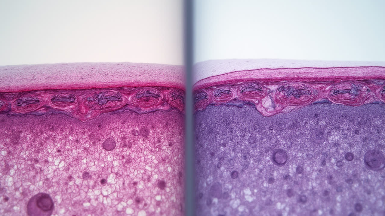
One of the most fascinating aspects of the basal lamina is its structural composition. When observed under an electron microscope (it's not visible with standard light microscopy), the basal lamina reveals two distinct layers: the lamina lucida and the lamina densa. The lamina lucida is an electron-lucent (appearing lighter) layer situated closest to the basal surface of cells, while the lamina densa appears as an electron-dense (darker) layer positioned slightly farther away. This dual-layer organization contributes to the basal lamina's functional versatility.
The molecular composition of basal lamina includes specific components dedicated to each layer. The lamina lucida contains glycoprotein laminin, a cross-shaped protein that helps connect cells to the extracellular matrix. Meanwhile, the lamina densa primarily consists of type IV collagen, which forms a mesh-like network providing structural support. Together, these molecules create a specialized environment that influences the behavior of adjacent cells.
Functions of Basal Lamina
The basal lamina performs several essential functions in the body's tissues. Its primary role is anchoring epithelial cells to the underlying extracellular matrix, providing stability and structural integrity. Without this secure attachment, tissues would lose their organization and function. Think of it as the biological equivalent of adhesive that holds different tissue components together.
Beyond mere attachment, the basal lamina actively influences the cells it contacts. It organizes proteins within adjacent cell membranes, creating specialized domains that affect cellular behavior. The basal lamina also induces cell differentiation and regulates metabolism, essentially providing instructions that help cells maintain their specialized functions. Isn't it remarkable how a seemingly simple layer can exert such profound control over cellular behavior?
Another critical function of the basal lamina is its role in cell migration. During development and tissue repair, cells often need to move through tissues in a controlled manner. The basal lamina serves as a highway of sorts, guiding cells along specific routes. Additionally, it helps establish and maintain cell polarity—the asymmetric organization of cellular components that allows specialized functions on different sides of the cell. This orientation is crucial for proper tissue functioning, especially in organs with directional functions like the intestines or kidneys.
What is Basement Membrane?
The basement membrane is a thin, sheet-like extracellular structure that forms an anatomical barrier where epithelial cells meet connective tissue. Unlike the basal lamina, the basement membrane is visible under light microscopy without special staining techniques. This greater visibility stems from its more substantial structure, which includes an additional layer beyond what's found in the basal lamina.
When examined under electron microscopy, the basement membrane reveals three distinct layers: lamina lucida, lamina densa, and lamina reticularis. The first two layers are identical to those found in the basal lamina, while the lamina reticularis is an electron-lucent layer unique to the basement membrane. This third layer contains anchoring fibrils and microfibrils that connect the basement membrane to the underlying connective tissue, providing additional structural support and integration with surrounding tissues.
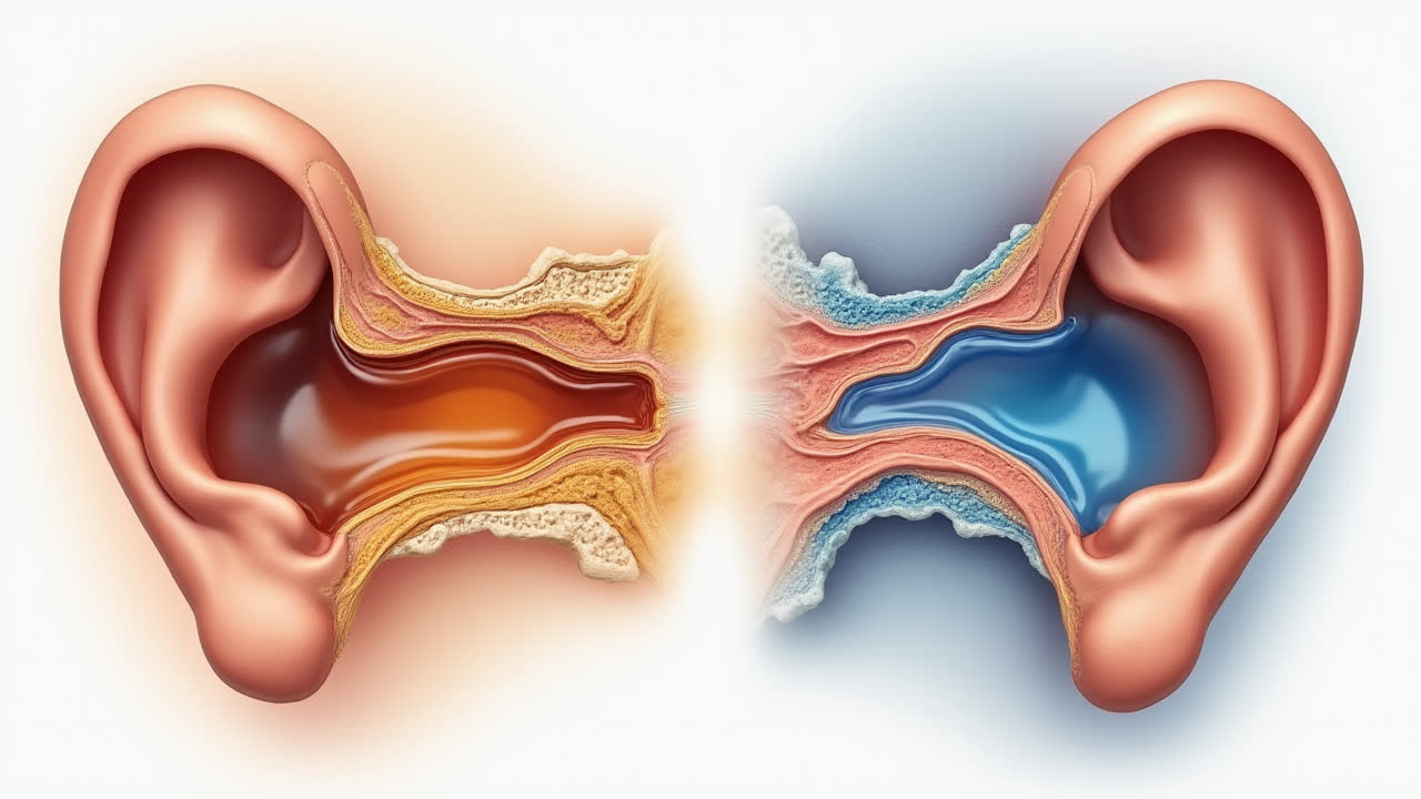
The basement membrane appears in various specialized forms throughout the body. Examples include the basilar membrane in the inner ear, Bruch's membrane in the eye, Descemet's membrane in the cornea, and the glomerular basement membrane in the kidneys. Each of these specialized versions has adaptations suited to its specific location and function, demonstrating the versatility of this fundamental structure in different organ systems.
Functions of Basement Membrane
The basement membrane serves numerous vital functions in the body. As an anatomical barrier, it provides a distinct boundary between epithelial cells and connective tissue, maintaining tissue architecture and organization. This separation is crucial for proper tissue function and prevents the inappropriate mixing of different cell types.
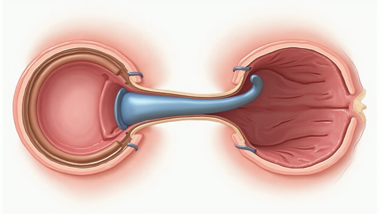
Different specialized basement membranes perform unique functions based on their location. For instance, the basilar membrane within the cochlea of the inner ear is a stiff structure that separates two liquid-filled tubes (the scala media and scala tympani). This membrane moves in response to sound waves, playing a crucial role in hearing. In contrast, Bruch's membrane in the eye provides structural support to the retinal pigment epithelium, contributing to proper vision. Who would have thought such a thin structure could play such diverse roles throughout the body?
Perhaps one of the most remarkable specialized basement membranes is the glomerular basement membrane found in kidneys. This structure represents a fusion of podocytes and endothelial basal lamina, creating a selective permeability barrier essential for urine production. It filters blood plasma, allowing water and small molecules to pass while retaining proteins and cells within the bloodstream—a sophisticated filtration system at the microscopic level. The basement membrane's ability to adapt to such specialized functions highlights its importance in organ-specific processes.
Comparing Basal Lamina and Basement Membrane
| Characteristic | Basal Lamina | Basement Membrane |
|---|---|---|
| Definition | Layer of extracellular matrix found on the basal surface of epithelial cells | Thin, dense layer of extracellular matrix that lines most human tissues |
| Secretion | Secreted by epithelial cells | Secreted by epithelial cells |
| Visibility | Only visible under electron microscope | Visible under light microscopy |
| Structure | Composed of two layers: lamina densa and lamina lucida | Composed of three layers: lamina densa, lamina lucida, and lamina reticularis |
| Components | Contains glycoprotein laminin and type IV collagen | Contains glycoprotein laminin, type IV collagen, and anchoring fibrils in lamina reticularis |
| Location | Found in epithelial tissue, fat cells, Schwann cells, and skeletal muscle fibers | Found where epithelial cells meet connective tissue and in specialized forms throughout the body |
| Primary Functions | Cell attachment, protein organization, cell differentiation, migration route | Anatomical barrier, structural support, specialized functions in different organs |
| Examples | Basal surface of epithelial cells, surrounding muscle fibers | Basilar membrane, Bruch's membrane, Descemet's membrane, Glomerular basement membrane |
Similarities Between Basal Lamina and Basement Membrane
Despite their differences, basal lamina and basement membrane share important similarities. Both are fundamental components of the extracellular matrix, forming specialized layers that interact with cells. These structures are primarily secreted by epithelial cells, highlighting their close relationship with epithelial tissues throughout the body.
Both structures serve as attachment points for epithelial cells, providing stability and facilitating communication between cells and their environment. They share common molecular components, including laminin and type IV collagen, which contribute to their structural integrity and functional capabilities. The presence of these similar proteins underscores the evolutionary relationship between these two extracellular structures.
Another similarity lies in their role in tissue organization. Both basal lamina and basement membrane help maintain proper tissue architecture by creating boundaries between different cell types and tissue compartments. They also influence cellular behavior, affecting processes like differentiation, migration, and metabolism. These shared functions reflect their fundamental importance in maintaining tissue integrity and facilitating proper organ function throughout the body.
Frequently Asked Questions
Can basal lamina exist without a basement membrane?
Yes, the basal lamina can exist without developing into a complete basement membrane. In some tissues, epithelial cells secrete only the components necessary for basal lamina formation (lamina densa and lamina lucida), without the additional lamina reticularis layer that would make it a basement membrane. This occurs in locations where the attachment to underlying connective tissue is less substantial or where the tissue's functional requirements don't necessitate the additional structural support provided by a full basement membrane.
What happens when basement membranes are damaged?
Damage to basement membranes can lead to several pathological conditions. When basement membranes are compromised, tissue integrity is affected, potentially leading to loss of organ function. For example, in diabetic nephropathy, the glomerular basement membrane in kidneys thickens abnormally, impairing filtration and potentially leading to kidney failure. In skin blistering diseases like epidermolysis bullosa, genetic defects in basement membrane components result in skin fragility and blister formation. Additionally, cancer cells must breach the basement membrane to invade surrounding tissues and metastasize, making basement membrane integrity crucial in cancer progression.
How do scientists study basal lamina and basement membranes?
Scientists employ several techniques to study these structures. Electron microscopy is essential for visualizing the basal lamina, while both light and electron microscopy can be used for basement membranes. Immunohistochemistry helps identify specific molecular components by using antibodies that target proteins like laminin or collagen IV. Cell culture systems, particularly those involving epithelial cells grown on artificial matrices, allow researchers to observe basement membrane formation in controlled environments. Genetic approaches, including knockout mouse models lacking specific basement membrane components, help determine the functional importance of individual proteins. These combined approaches have significantly advanced our understanding of these crucial extracellular structures.
Conclusion
The basal lamina and basement membrane, while similar in many respects, represent distinct structures with important differences in composition, visibility, and function. The basal lamina consists of two layers—lamina densa and lamina lucida—and is only visible under electron microscopy. In contrast, the basement membrane includes these two layers plus the lamina reticularis, making it more substantial and visible even under light microscopy.
Understanding the distinction between these structures is not merely an academic exercise but has practical implications for medicine and biology. Many pathological conditions involve abnormalities in these extracellular components, including kidney diseases, skin blistering disorders, and cancer metastasis. By appreciating the unique properties of basal lamina and basement membrane, researchers and clinicians can better understand tissue organization in health and disease.
As our technological capabilities advance, we continue to uncover new details about these fascinating structures and their roles in normal physiology and disease processes. From development to aging, basal lamina and basement membrane remain integral to our understanding of how tissues maintain their structure and function throughout life. The next time you encounter these terms, you'll appreciate the subtle but significant differences that make each unique in the complex world of cellular biology.